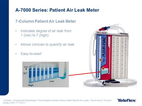
Chest Tube Air Leak

Chest tube air leaks are a critical concern in medical practice, particularly in the context of thoracic surgery and trauma care. An air leak occurs when air escapes from the lung and passes through a chest tube inserted to manage pneumothorax or other lung-related injuries. These leaks can complicate recovery and require careful management to ensure patient safety and optimize outcomes. In this comprehensive guide, we will delve into the intricacies of chest tube air leaks, exploring their causes, clinical implications, management strategies, and the latest advancements in this field.
Understanding Chest Tube Air Leaks

Chest tube air leaks are a common complication encountered in thoracic surgery and critical care settings. These leaks occur when air escapes from the lung parenchyma or pleural space and passes through the chest tube drainage system. The presence of an air leak can indicate an underlying lung injury or pathology, such as a pneumothorax, bronchopleural fistula, or post-surgical complications.
The management of chest tube air leaks is crucial to prevent further lung damage, control air exchange, and promote healing. Timely recognition and appropriate intervention are essential to ensure patient comfort and facilitate recovery. This section provides an in-depth understanding of chest tube air leaks, including their causes, clinical presentation, and the impact on patient care.
Causes of Chest Tube Air Leaks
Chest tube air leaks can arise from various etiologies, each presenting unique challenges in management. The primary causes of air leaks include:
- Traumatic Pneumothorax: Chest tube air leaks frequently occur in the setting of traumatic injuries, such as blunt or penetrating chest trauma. These injuries can lead to the development of a pneumothorax, where air accumulates in the pleural space, requiring chest tube insertion for drainage.
- Iatrogenic Injuries: Medical procedures, including thoracic surgery, biopsy, or catheterization, can inadvertently cause lung injuries resulting in air leaks. Proper technique and careful monitoring are essential to minimize the risk of iatrogenic air leaks.
- Post-Surgical Complications: Air leaks may occur as a complication of thoracic surgery, particularly following procedures involving lung resection or lung transplant. These leaks can be a result of bronchopleural fistula formation or tissue damage during surgery.
- Spontaneous Pneumothorax: In some cases, chest tube air leaks can arise from spontaneous pneumothorax, a condition where air leaks into the pleural space without an obvious precipitating event. This can be associated with underlying lung diseases or congenital anomalies.
Clinical Presentation and Diagnosis
The clinical presentation of chest tube air leaks can vary depending on the underlying cause and the severity of the leak. Common signs and symptoms include:
- Audible Air Leak: A high-pitched or bubbling sound from the chest tube drainage system, often described as a "squeak" or "sizzle," is a characteristic finding in air leaks.
- Decreased Lung Sounds: Auscultation of the lung fields may reveal decreased or absent breath sounds over the affected lung, indicating a possible air leak.
- Subcutaneous Emphysema: Air tracking along the chest wall and subcutaneous tissues, leading to visible crepitus, can be a sign of an extensive air leak.
- Chest Tube Output: Increased or continuous air output from the chest tube, especially in the absence of significant fluid drainage, is suggestive of an air leak.
- Dyspnea and Tachypnea: Patients may experience shortness of breath and rapid breathing, especially with larger air leaks affecting lung function.
Diagnosing chest tube air leaks involves a combination of clinical assessment, chest radiography, and sometimes advanced imaging techniques. Chest X-rays are essential to evaluate the presence and extent of pneumothorax and identify any associated lung injuries. Computed tomography (CT) scans may be indicated in complex cases to provide detailed anatomical information.
Management Strategies
The management of chest tube air leaks aims to control the leak, prevent further lung damage, and promote healing. The specific approach depends on the underlying cause, the severity of the leak, and the patient’s overall clinical condition. Here are some key management strategies:
- Water Seal Chamber: Chest tubes are typically connected to a water seal chamber, which acts as a one-way valve, allowing air to escape but preventing its re-entry. This system helps to control the air leak and maintain a negative pressure environment within the pleural space.
- Suction Control: Adjusting the suction pressure applied to the chest tube can influence the management of air leaks. Lower suction levels may be used initially to minimize trauma to the lung tissue, while higher suction may be required to control more significant leaks.
- Chest Tube Positioning: Optimal chest tube positioning is crucial to ensure effective drainage and minimize the risk of air leaks. Chest tubes should be positioned in the pleural space, avoiding lung parenchyma, to prevent tissue injury and promote drainage.
- Chest Tube Clamping: In certain cases, clamping the chest tube for a short duration may be considered to assess the leak's nature and determine the appropriate management strategy. Clamping should be done under close monitoring to avoid the risk of tension pneumothorax.
- Additional Chest Tubes: If a single chest tube is insufficient to control the air leak, additional tubes may be inserted to enhance drainage and manage the leak effectively. The number and position of chest tubes depend on the patient's anatomy and the nature of the leak.
- Bronchopleural Fistula Management: In cases of bronchopleural fistula, where air leaks from the bronchus into the pleural space, specific interventions may be required. These can include bronchoscopic techniques, such as stenting or fibrin glue application, to seal the fistula and control the air leak.
Advancements in Chest Tube Air Leak Management

The field of chest tube air leak management has seen significant advancements in recent years, driven by technological innovations and a better understanding of lung physiology. These advancements aim to improve patient outcomes, reduce the duration of chest tube drainage, and minimize complications associated with air leaks.
Innovative Chest Tube Systems
Traditional chest tubes, often made of silicone or polyvinyl chloride (PVC), have been the mainstay of chest drainage for decades. However, recent advancements have led to the development of more sophisticated chest tube systems designed to enhance leak management and patient comfort.
One notable innovation is the introduction of active chest drainage systems, which actively monitor and regulate the pressure within the pleural space. These systems utilize advanced sensors and microprocessors to continuously measure and control the negative pressure, ensuring optimal drainage while minimizing the risk of excessive suction and tissue trauma.
Additionally, miniaturized chest tubes have gained popularity, particularly in pediatric and minimally invasive surgical settings. These smaller-diameter tubes offer improved patient comfort, reduced tissue trauma, and easier insertion, especially in patients with narrow intercostal spaces or delicate lung tissue.
Enhanced Imaging Techniques
Advanced imaging modalities have revolutionized the diagnosis and management of chest tube air leaks. High-resolution computed tomography (HRCT) scans provide detailed anatomical information, allowing for precise identification of the leak’s origin and extent. This advanced imaging helps guide targeted interventions and improves patient selection for specific treatment approaches.
Furthermore, dynamic CT imaging, which captures the lung's movement during ventilation, has emerged as a valuable tool in evaluating chest tube air leaks. By visualizing the dynamic behavior of the lung and chest tube system, physicians can better understand the leak's mechanism and tailor management strategies accordingly.
Biomaterial-Based Interventions
The use of biomaterials and tissue adhesives has shown promise in managing chest tube air leaks, particularly in cases of bronchopleural fistula. Fibrin sealants and glues, derived from human plasma, have been successfully employed to seal leaks and promote healing. These materials mimic the body’s natural healing process, providing a scaffold for tissue regeneration and enhancing the sealing of air leaks.
Additionally, the development of bioactive scaffolds and hydrogels holds great potential for future air leak management. These biomaterials can be engineered to deliver therapeutic agents, such as growth factors or antibiotics, directly to the site of injury, promoting tissue regeneration and reducing the risk of infection.
Clinical Implications and Patient Outcomes
The timely and effective management of chest tube air leaks is crucial for optimizing patient outcomes and minimizing complications. Delayed or inadequate management can lead to prolonged chest tube drainage, increased risk of infection, and potential lung damage.
Prolonged chest tube drainage, particularly in the presence of air leaks, can result in prolonged hospital stays and increased healthcare costs. Additionally, the risk of infectious complications, such as empyema or pneumonia, is heightened with persistent air leaks, as the chest tube provides a potential route for bacterial entry. Proper leak management and early chest tube removal are essential to prevent these complications.
Moreover, chest tube air leaks can impact lung function and respiratory mechanics. Persistent air leaks can lead to atelectasis, lung collapse, or impaired gas exchange, affecting the patient's respiratory status and overall clinical condition. Effective leak management, including the use of advanced chest tube systems and targeted interventions, can help preserve lung function and promote optimal recovery.
Future Directions and Research
The field of chest tube air leak management continues to evolve, driven by ongoing research and technological advancements. Several areas of future research and development hold promise for further improving patient care and outcomes.
Smart Chest Tube Systems
The development of smart chest tube systems equipped with advanced sensors and real-time monitoring capabilities is an exciting prospect. These systems can continuously assess chest tube function, detect air leaks, and provide real-time feedback to healthcare providers. By integrating artificial intelligence and machine learning algorithms, smart chest tubes can optimize leak management, reduce the need for frequent chest X-rays, and enhance patient safety.
Personalized Treatment Approaches
Personalized medicine, tailored to individual patient characteristics and needs, is gaining traction in various medical specialties. In the context of chest tube air leak management, personalized approaches can involve tailoring chest tube selection, suction levels, and management strategies based on patient-specific factors such as age, lung function, and comorbidities. This patient-centric approach has the potential to improve outcomes and reduce adverse events.
Biomarker Discovery
The identification of specific biomarkers associated with chest tube air leaks and their resolution can provide valuable insights into the underlying pathophysiology and guide targeted interventions. Ongoing research is focused on discovering and validating biomarkers that can predict the risk of air leaks, monitor their resolution, and assess the effectiveness of therapeutic interventions.
Conclusion

Chest tube air leaks are a complex and challenging complication in thoracic surgery and critical care. However, with advancements in chest tube technology, imaging techniques, and biomaterial-based interventions, healthcare providers now have a wider range of tools and strategies to manage these leaks effectively. The timely recognition and appropriate management of chest tube air leaks are essential to ensure patient comfort, promote healing, and optimize clinical outcomes.
As the field continues to evolve, ongoing research and innovation will further enhance our understanding of chest tube air leaks and drive the development of novel management approaches. The integration of smart technologies, personalized medicine, and biomarker-guided therapies holds great promise for improving patient care and outcomes in the management of chest tube air leaks.
What are the common causes of chest tube air leaks?
+Chest tube air leaks can arise from traumatic injuries, iatrogenic procedures, post-surgical complications, and spontaneous pneumothorax. The specific cause depends on the patient’s clinical history and underlying lung pathology.
How are chest tube air leaks diagnosed?
+Chest tube air leaks are diagnosed through a combination of clinical assessment, chest X-rays, and sometimes advanced imaging techniques like CT scans. Auscultation of lung sounds, audible air leaks, and changes in chest tube output are key indicators.
What are the management strategies for chest tube air leaks?
+Management strategies include water seal chambers, suction control, chest tube positioning, and additional chest tubes. Bronchopleural fistula may require specific interventions like bronchoscopic techniques or fibrin glue application.
How do advanced chest tube systems improve leak management?
+Advanced chest tube systems, such as active chest drainage systems, utilize sensors and microprocessors to regulate pressure and optimize drainage. Miniaturized chest tubes offer improved patient comfort and reduced tissue trauma.
What are the future directions in chest tube air leak management research?
+Future research focuses on smart chest tube systems with real-time monitoring, personalized treatment approaches, and biomarker discovery. These advancements aim to improve patient outcomes and reduce complications.


